Press Releases
JAMSTEC
Kochi University
National Agriculture and Food Research Organization
Japan Synchrotron Radiation Research Institute
Ehime University
Hiroshima University
High Energy Accelerator Research Organization
The University of Tokyo
Discovery of abundant ferromanganese microparticles in oxic pelagic sediments:
New insights into the global budget of metallic elements
Overview
A research group led by Drs. Yuki Morono and Fumio Inagaki at the Japan Agency for Marine-Earth Science and Technology (JAMSTEC) and Dr. Go-Ichiro Uramoto at Kochi University have investigated subseafloor sediments in the ultra-oligotrophic region of the South Pacific Gyre (SPG) (Figure 1). By using high-resolution imaging techniques, they found abundant (108–109 particles/cm3) micrometer-scale ferromanganese mineral particles (Mn-microparticles) in the oxic pelagic clays of the SPG (Figure 2). Major and trace element compositional analyses revealed that iron and manganese were the major components of Mn-microparticles, while they also contained rare-earth elements*1. From the number of Mn-microparticles per unit volume of sediment and the global distribution of oxic pelagic clays, they estimated the number of Mn-microparticles to be 1.5–8.8 × 1028 particles, which accounted for 1.28–7.62 Tt of manganese. This estimate is at least 2 orders of magnitude larger than the manganese budget for manganese nodules and manganese crusts on the seafloor. Additionally, they also found that these Mn-microparticles contained 3.3–19.4 Gt of rare-earth elements and that subseafloor Mn-microparticles contribute significantly to the global budget of metallic elements.
Manganese is the third most abundant metallic element, after iron and titanium, in the Earth’s crust, and ferromanganese minerals are sensitive to changes in oxidation state. Assessing the formation and preservation of ferromanganese minerals is important for understanding the global marine cycles of manganese and numerous associated trace elements. The most extensive manganese mineral deposits occur on abyssal plains, including those below open-ocean gyres. However, the extent of ferromanganese minerals buried in subseafloor sediments remains unclear.
In 2010, the Integrated Ocean Drilling Program (IODP)*2 Expedition 329 drilled the seafloor in the ultra-oligotrophic SPG to investigate the deep-sea sedimentary environment (Figure 1) (as reported on October 8, 2010). Geochemical and microbiological studies of sediment core samples*3 from six SPG drill sites at water depths of 3740–5695 m revealed the presence of dissolved oxygen and aerobic microbial communities throughout the sedimentary sequence. Moreover, these characteristics extended from the present-day to the mid-Cretaceous sedimentary layer above the crustal basement (as reported on March 17, 2015). These oxic sediments represent up to ~44% of the Pacific Ocean and ~37% of the global ocean (Figure 1).
To better characterize micrometer-scale sedimentary microstructures in the entirely oxic SPG sediments, JAMSTEC and Kochi University researchers investigated the ultra-fine structures of marine sediment core samples of various oxidation states, ranging from continental margins to an open-ocean gyre.
The researchers found a remarkable number of Mn-microparticles (108–109 particles/cm3), but only in oxic pelagic clays. From the global distribution of oxic pelagic clays, they estimated the total number of these Mn-microparticles to be 1.5–8.8 × 1028 particles within the global ocean. Additionally, they developed a new technique for separating Mn-microparticles from sedimentary matrices by combining a sequential density concentration*4 and flow cytometry/particle sorting*5, which enabled in-depth analyses of the texture and major and trace element compositions of Mn-microparticles. Through 2D and 3D morphological characterization, they demonstrated the presence of a clump of tangled, fibrous strands of ferromanganese minerals in the Mn-microparticles. The Mn-microparticles contained particularly high amounts of manganese, which accounted for a mean 42% of the total manganese in the oxic sediment samples (Figure 4).
The total amount of manganese contained in Mn-microparticles is estimated to be 1.28–7.62 Tt in the global ocean, which is at least 2–3 orders of magnitude higher than those presented in previous studies based on seafloor manganese nodules and manganese crusts, in which the proposed budgets of manganese have ranged from 0.706–2.60 Gt (Figure 4). Mn-microparticles are also estimated to harbor 3.3–19.4 Gt of rare-earth elements globally. Their major and trace element compositions suggest that these particles were mainly precipitated from bottom waters. The flux of particle sedimentation was found from their abundance in the sediments to be ~100 particles/cm2 per day into oxic sediments. Along with the examination of manganese input, formation, and preservation, this study provides new insights into the global budget of metallic elements in the abundant Mn-microparticles present in deep subseafloor environments.
The samples used in this study were collected during IODP Expedition 329, “South Pacific Gyre Subseafloor Life.” Three-dimensional imaging of Mn-microparticles was performed using high-resolution X-ray microtomography at the beamlines BL20B2, BL20XU, and BL47XU of the Japan Synchrotron Radiation Research Institute and X-ray absorption spectral analyses were performed by scanning transmission X-ray microscopy (STXM) and using an X-ray absorption fine structure (XAFS) facility at the beamlines BL-9A and BL13A of the Photon Factory of the High Energy Accelerator Research Organization.
This study was supported in part by the Japan Society for the Promotion of Science (JSPS) Strategic Fund for Strengthening Leading-Edge Research and Development, the JSPS Funding Program for Next Generation World-Leading Researchers (GR102), JSPS Grant-in-Aid for Scientific Research (no. 24687004, 25871219, 26251041, 14J00199, 15H02810, 15H05608, 17H04582, 17H06458, 18H04134), and Ministry of Education, Culture, Sports, Science, and Technology (MEXT) Fund Leading Initiative for Excellent Young Researchers.
The above results were published in Nature Communications on February 6, 2019 (JST).
Title: Significant contribution of subseafloor microparticles to the global manganese budget
Authors: Go-Ichiro Uramoto1,2, Yuki Morono1,3, Naotaka Tomioka1, Shigeyuki Wakaki1, Ryoichi Nakada1, Rota Wagai4, Kentaro Uesugi5, Akihisa Takeuchi5, Masato Hoshino5, Yoshio Suzuki5,6, Fumito Shiraishi7, Satoshi Mitsunobu8, Hiroki Suga7,12, Yasuo Takeichi9, Yoshio Takahashi10 & Fumio Inagaki1,3,11
Affiliations: 1. Kochi Institute for Core Sample Research, JAMSTEC 2. Center for Advanced Marine Core Research, Kochi University 3. Research and Development Center for Submarine Resources, JAMSTEC 4. Institute for Agro-Environmental Sciences, National Agriculture and Food Research Organization 5. Japan Synchrotron Radiation Research Institute 6. Graduate School of Frontier Sciences, The University of Tokyo 7. Department of Earth and Planetary Systems Science, Hiroshima University 8. Department of Environmental Conservation, Graduate School of Agriculture, Ehime University 9. Institute of Materials Structure Science, High Energy Accelerator Research Organization 10. Department of Earth and Planetary Science, The University of Tokyo 11. Research and Development Center for Ocean Drilling Science, JAMSTEC 12. Present address: Department of Earth and Planetary Science, The University of Tokyo.
*1 Rare-earth elements are a set of seventeen chemical elements in the periodic table, including the fifteen lanthanides, scandium, and yttrium. These elements are all metals that are very difficult to mine because it is unusual to find them in concentrations high enough for economical extraction. They have unique magnetic or optical properties, and thus are important in the construction of many everyday devices, such as computer memory chips, rechargeable batteries, cell phones, magnets, fluorescent lighting, and much more.
*2 The Integrated Ocean Drilling Program (IODP) was a multinational cooperative project that ran from 2003 to 2013 under the initiatives of Japan, the United States, and the European countries. The scientific drilling vessels D/V Chikyu operated by Japan and the JOIDES Resolution by the U.S, and the option to charter mission-specific platforms by Europe were utilized for expeditions. The research was aimed at shedding light on global environmental changes, the Earth’s mantle and crustal dynamics and tectonics, and the biosphere beneath the seafloor. Since October of 2013, the project has been operated under a new framework as the International Ocean Discovery Program (IODP).
*3 Core samples are columnar samples of sediments (or rocks) collected by drilling. JAMSTEC and Kochi University jointly operate the Kochi Core Center (Nankoku City, Kochi), where core samples (with total lengths reaching 130 kilometers) collected from the western Pacific, Indian Ocean, and other seas via ocean drilling are stored.
*4 Density concentration is a sample processing technique for mineral materials based on the fact that different minerals exhibit different densities. If a mixture of minerals with different densities is suspended in a heavy metal solution of an intermediate density, the grains with densities less than that of the solution will float and grains with densities greater than the solution will sink. This study revealed the density of Mn-microparticles and used this technique as a pre-processing protocol of the flow cytometer/particle sorting technique.
*5 Flow cytometry/particle sorting is a laser-based method that is used to separate mineral particles from sediment samples on the basis of their size and fluorescence intensity. Flow cytometry was originally used to count and sort biological cells. Cells in a stream of liquid passing an irradiating laser are separated on the basis of their size, fluorescence intensity, and wavelength. In this study, the researchers succeeded in applying this biology-based technique for Mn-microparticle separation by identifying their unique optical characteristics, including scattering and fluorescence.
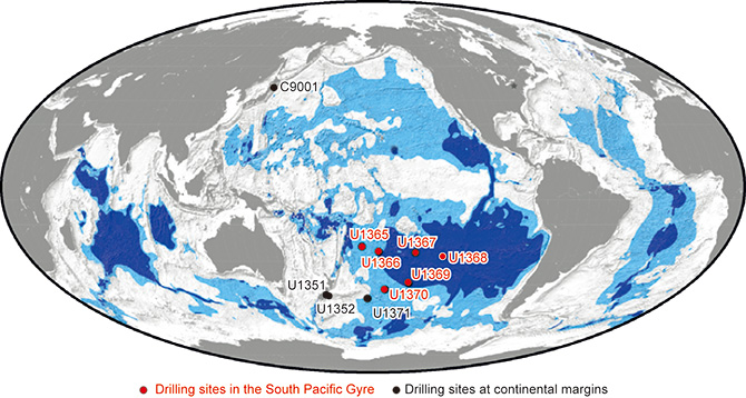
Figure 1 Locations of sampling sites. Sites are plotted on a global map showing regions that may contain dissolved oxygen and aerobic activity through the entire sedimentary sequence. Dark (light) blue indicates regions likely to feature maximum (minimum) dissolved oxygen and aerobic activity.
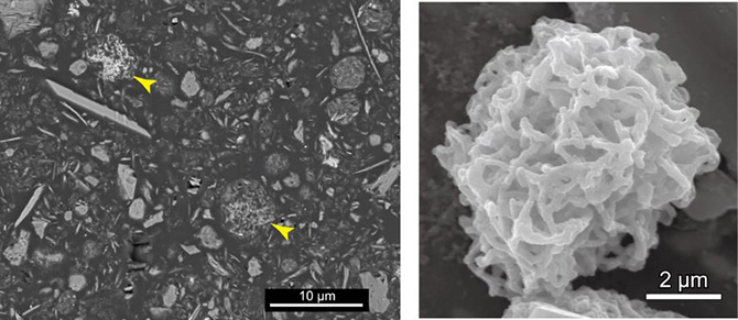
Figure 2 (Left) Cross-sectional scanning electron microscopy (SEM) images of resin-embedded oxic pelagic clay. Arrows indicate Mn-microparticles (yellow); (right) SEM image of a Mn-microparticle in a density-separated sample.
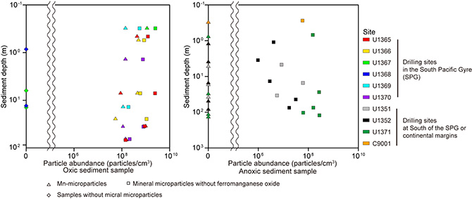
Figure 3 (Left) Depth profiles of the number of Mn-microparticles in pelagic oxic sediment samples from the South Pacific Gyre; (right) depth profiles of the number of clay microparticles in anoxic sediment samples from the South Pacific and continental margins.
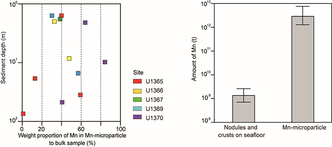
Figure 4 (Left) Depth profiles of the proportion of Mn-microparticle manganese mass to bulk sample mass (%); (right) comparison of the manganese budgets estimated for Mn-nodules and crusts on the seafloor and Mn-microparticles in oxic pelagic clay. Whiskers represent maximum and minimum estimates.
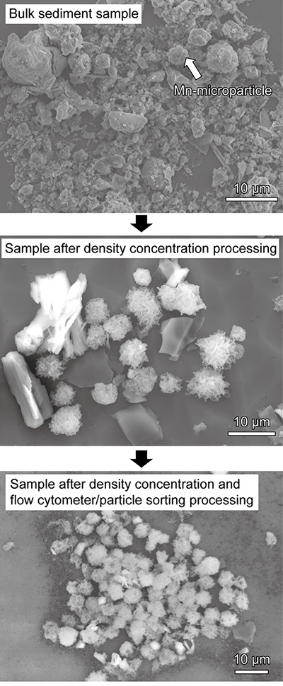
Figure 5 SEM images showing the Mn-microparticle concentration and separation results. (Upper) Bulk sediment sample; (middle) sample after density concentration processing; (lower) sample after density concentration and flow cytometry/particle sorting.
Contacts:
- (For this study)
- Yuki Morono, Deputy Group Leader, Geomicrobiology Group, Kochi Institute for Core Sample Research, JAMSTEC
- Go-Ichiro Uramoto, Specially Appointed Assistant Professor, Center for Advanced Marine Core Research, Kochi University
- Yoshio Takahashi, Professor, Department of Earth and Planetary Science,
The University of Tokyo - (For press release)
- Tsuyoshi Noguchi, Manager, Press Division, Public Relations, JAMSTEC
- Hikino Hajime, KEK PR Office, HIGH ENERGY ACCELERATOR RESEARCH ORGANIZATION
- Kristina Awatsu, Office of Communication, Graduate School of Science,
The University of Tokyo