Press Releases
JAMSTEC
Untangling the family-lineages of calcifying red algae
Nanoscale crystals reflect genetic relationships of often overlooked ecosystem building seaweeds.
1. Key Points
- ♦
- Until now the morphologic classification of calcifying red algae – important ecosystem engineers in shallow marine environments – was difficult, because it was very different from the phylogenetic classification base on DNA-sequences.
- ♦
- New research shows that nanocrystals in the skeleton of calcifying red algae reflect their genetic relationships.
- ♦
- Using this ultrastructure for classification also provides new ways to link genetic adaption and evolution of organisms with their morphological adaption.
- ♦
- These results are expected to provide new research avenues to better assess the impact of global warming and ocean acidification on sensitive shallow marine and coastal ecosystems.
2.Background
Coralline red algae are a group of usually reddish or pinkish seaweeds that are able to build hard skeletons made out of the mineral calcite. These calcified red algae are common sights along the shores of the world (Fig 1a,b) and can most often be recognized as pink and red crusts on rock, embankments and old seashells. They also live solitary as “living rocks” called Rhodoliths or Maerl. A close look at almost any rocky shore or coral reef will reveal an abundance of pink to pinkish-grey patches, splashed as though by a mad painter over every rock surface (Fig 1c). In coral reefs, they act as the glue holding the reefs together, and play a major role in the stabilization, initiation and subsequent growth of reefs. Sometimes especially in temperate and polar regions these calcifying algae even form their own unique ecosystems called “Maerl beds”, where coralline red algae cover the ocean floor completely (Fig. 1a). These Maerls form the basis of marine life where they occur, offering physical refuge and protection from predation as well as productive feeding grounds for fish. Maerl also acts as nurseries for many commercially important fish and shellfish species. Because of that coralline red algae are often considered as ‘ecosystem engineers’ that not only play a vital role in reef development but virtually every shallow marine environment. However, despite their ubiquitous occurrence and important role in many ecosystems, coralline red algae were mostly overlooked in the past. They suffered this fate mainly due to the fact that these unique organisms have a complex history of evolution and species are often hard to reliably identify, making them difficult to study in detail. Often the classification of coralline algal species differs completely, depending on if it is based on either DNA sequencing or morphological studies. These difficulties led to a state, where, despite their obvious importance to marine life, coralline red algae are often simply ignored. In particular, their role in ecosystems but also their response to ongoing climate change and the resulting effect on ecosystems is still largely unknown.
This is essential, since these environments were coralline red algae live, are under threat by modern climate change. Rising temperatures and ocean acidification are a major and concerning effect of climate change as they may change or even inhibit the way organisms such as corals, clams and algae produce their skeletons and thereby “build” the foundations of coral reefs and other marine ecosystems. Consequently, understanding how this “biomineralization” works and especially it is affected by climate change is becoming more and more. Interestingly, the way coralline red algae form their calcite skeleton is a completely unique feature in nature. They are the only known living organisms that deposit calcium carbonate within the organic walls of individual cells (Fig. 2). This calcite formation is biochemically controlled and produces mineralization along microscopic organic strands made of cellulose and other compounds. These so-called polysaccharide microfibrils weave together to from the organic cell wall of red algae but are also found in many other organisms. Although the general organization of cells is similar for all organisms, the specific structure of this “polysaccharide fabric” and the chemical “microenvironments” the living cells generate within them, appear to be unique for different species. Thus, the mineral skeletons formed within these cellular microenvironments can be very different for various species, even within the same organism group.
In this study, we present a new way to classify coralline red algae by visualizing the differences in nanometer sized crystals that are found within their unique calcite skeleton: so-called “skeletal ultrastructures”.
Title: Nanocrystals as phenotypic expression of genotypes— an example in coralline red algae
Authors: Gerald Auer1,2, Werner E. Piller2
1. Research Institute for Marine Resources Utilization, Biogeochemistry Program, Japan Agency for Marine-Earth Science and Technology
2. University of Graz, Austria
3.Results
The authors use data from a collection of globally distributed red algae species (Fig. 3). They the shape of individual crystals that form in the cell walls of so called “epithallial” cells forming a skin-like layer at the outside of the tissue forming the body of red algae called the “thallus”.
The crystal forms found in the cell wall of these epithallial cells are unique within each group of coralline red algae (Fig. 4). Since these crystals are present on all calcified cells, they are simple to study and can be easily described, making them a potentially useful tool for taxonomy, the branch of science dealing with the classification of organisms (Fig. 4).
By combing these crystal shapes with other basic morphological characteristics such as the shape and organization of their reproductive organs, a taxonomic “family tree” of the species relationships was constructed (Fig. 4). This morphological phylogenetic tree describes the evolutionary relationships between the studied coralline red algal species. which coincides with phylogenies of coralline red algae based on the analysis of DNA sequences based on a study by Bittner et al. (2011) (Fig. 5). This overlap shows that genetic information on the relationships of these coralline red algae families is also expressed in the shape of the nanometer sized crystals forming their skeleton. This shows, that the right combination of descriptive micro- and/or nanoscale shapes in an organism can bridge the fields of organismal and molecular biology. Our new morphology, for the first time, allows the fast and reliable identification of calcifying red algae without the need for DNA sequencing.
4.Outlook:
This allows to study these often-overlooked organisms in our oceans and help us to fully understand them and their impact on the environment. Only by being able to easily identify which species live where we will be able to understand the role of individual coralline red algae have on various ecosystems. Understanding how skeletal shape and thus environmental adaption relates to genetic variability, also will allow us to gage the response of each species to changing environmental conditions. This will help to understand the effect climate change has on these vital but nearly unknown organisms in our oceans in order to protect them.
This study is however only a first step. Much more work will be needed to fully understand the impact and use these ultrastructures have in the evolutionary as well as the environmental adaption of organisms. Only by understanding these interactions can we start to fully understand how organisms evolved and adapted their skeletons to changing environments.
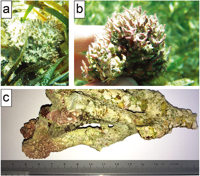
Figure 1: Images of coralline red algae used for this study. a) Free living red algae called a “rhodolith” from the sea grass meadows along the coast of Rodriguez Key in Florida (USA). b) close up of a rodolith. c) a rock covered by red algae and other sea weeds (Sesoko Jima, Okinawa, Japan).
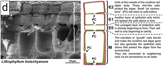
Figure 2: Electron microscopic picture of the body (= “thallus”) of a coralline red algae showing the outermost skin-like “epithallial” cells (E) and “growth” or “meristem” cells (M) that divide and extend the thallus below, making the algae grow. The organic “inside” of the cells are were lost during preparation, leaving only the calcified cell walls‚. Next to the image we show a cartoon explaining the function of these cells within the thallus.
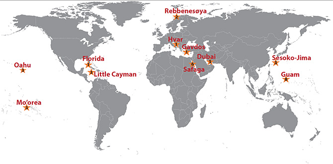
Figure 3: Map of the globally distributed samples used for this study shown as stars. Many samples are from the collection of the authors W. Piller and were taken during campaigns in the 1980s and 1990s. The numbers inside the stars describe the number of species that were used from each location.
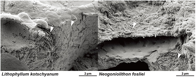
Figure 4: High magnification scanning electron microscope images showing the crystal ultrastructures found in the cell-wall of two species of coralline red algae Lithophyllum kotschyanum and Neogoniolithon fosliei. Note the size of the individual elements of only several hundred nanometers, and their distinct shape (white arrows).
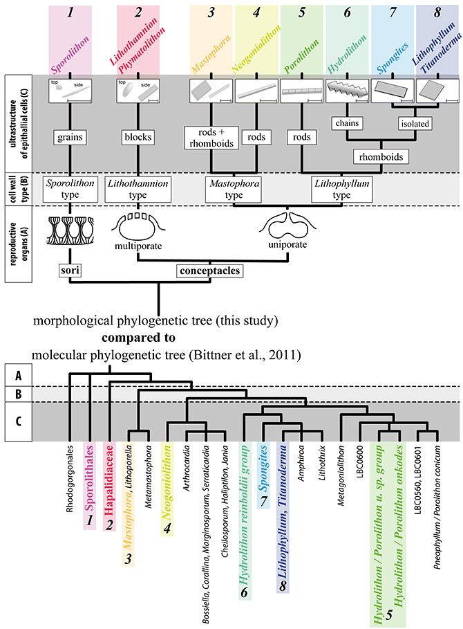
Figure 5: Phylogenetic tree showing the diagnostic position of the major morphological features used in the proposed taxonomical key. The proposed tree is consistent with the molecular phylogeny and allows the classification of all major groups of CRA based on three easily identifiable morphological features: A) reproductive organs, B) secondary cell-wall structures, and C) the dominant morphology of epithallial cell nanocrystals.
Contacts:
- (For this study)
- Gerald Auer, Young Research Fellow, Research Institute for Marine Resources Utilization, JAMSTEC
- (For press release)
- Public Relations Section, Marine Science and Technology Strategy Department, JAMSTEC